Hi Everyone, thanks for adding me to this group! I look forward to learning more about this condition and jugular compression, which I might have. Here’s a short version of my story. I’ve had neurological symptoms for over 12 years - headaches, speech issues (slurred words, non fluid speech), brain fog, anxiety, etc. Every neuro I saw couldn’t find anything and chalked it up to migraines or silent-migraines. I learned to live with it, but 2 years ago I developed and inner condition called semicircular canal dehiscence, which basically means that my skull base has thinned over time exposing a small part of the canal in my brain cavity. This comes with other challenging symptoms like ear fullness, sound sensitivity, tinnitus, etc. I’m considering a surgical option for this. A few docs also speculated with me that high head pressure might cause this condition over time. I’ve had lots of scans done over the last 2 years, but the radiology reports always said everything looked fine, other than my ears. However, I started loading some of my images in ChatGpt and it kept telling me that I have a moderate to severe compression of my left jugular vein, which looks apparent to a layman like. Anyway, that has led me to here so I can educate and advocate for myself meeting with doctors going forward. Thanks!
One other thing - is it okay to add images to posts? I would love feedback from more experienced members on my images, mainly to double check that I’m barking up the right tree. Thanks!
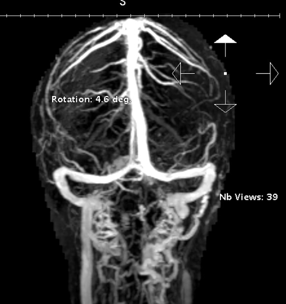


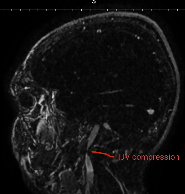
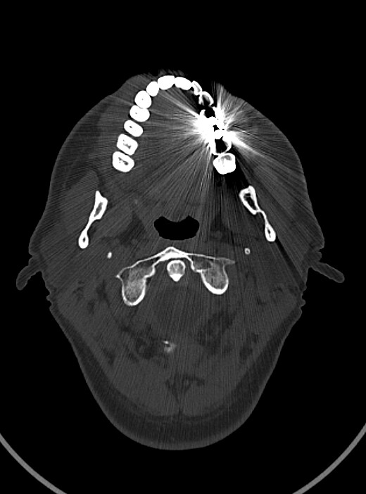
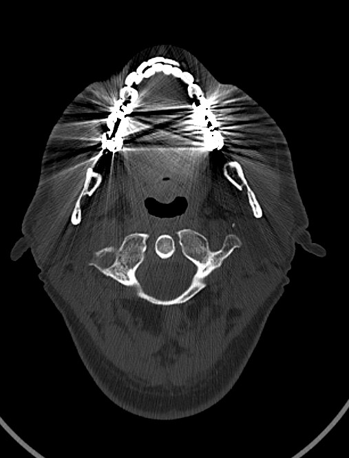
@tguerin18 did you happen to get a head/neck CT done? They are the best to look at vascular compression. If you did get one done, can you go to the axial view and go to C1? I attached my own imaging to help you know what to look for. Im less familiar with MRV, but I annotated your imaging. If I’m correct, your IJV is being compressed against your C1. Not sure if styloid is involved in the pinch or not. If you did get a CT done we will get to the bottom of it.
Thanks for the response and looking at my images. I have had several CTs of the head since that’s how they evaluate SSCD. Let me see if I can replicate your image. Was your CT with contrast?
I didn’t have contrast on my scan. Also, my image looks at different level than yours, so not sure if this is correct.
@tguerin18 this is pretty close, can you go down like 1 or two more slices so you can see the full wings of C1? I dont think I’ll need it to have contrast to guess what’s going on. Contrast would be best but I know my landmarks

@tguerin18 - The fact that your IJV completely disappears at the greatest point of compression indicates yours is significant. I agree w/ @TML that it appears to be at the level of C1. Our experience on this forum indicates that the IJV is often compressed between C1 & an elongated styloid process but there are also soft tissues such as muscle, nerve, other veins/arteries, lymph nodes, fascia or scar tissue that can also contribute to vascular compression. A CT scan w/ contrast of the area between your skull base & hyoid bone would be the best to help us (& you) see what’s going on.
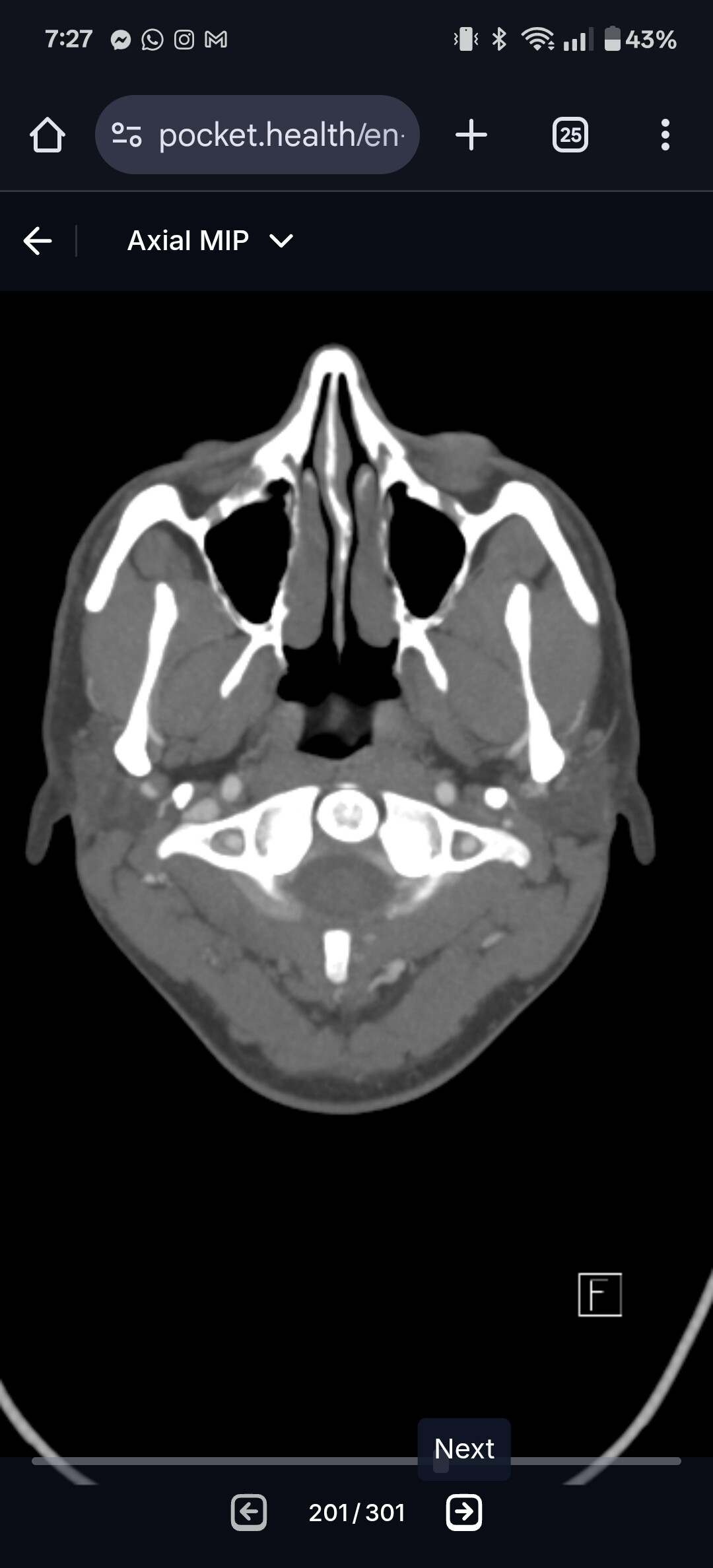
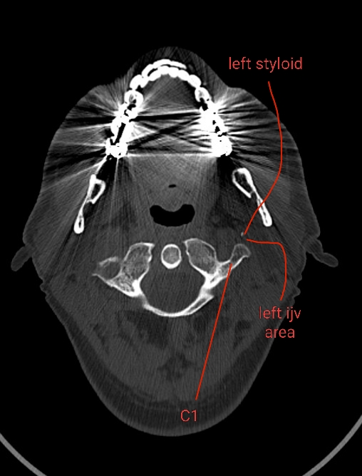
@tguerin18 see attached the annotated imaging. It appears your left ijv (right side of image) is compressed between your left styloid and C1. Since there is no matching white dot on the left side of the image, i suspect your left styloid is longer than your right and has caused a choke point for your left ijv between the styloid and C1. Since you don’t have contrast, I can’t say with 100% certainty the ijv is being compressed, but it is where the ijv is typically located.
Ijv compression can result in headaches, brain fog, memory difficulties, sleep difficulties, eye/ear pressure.
The vagus nerve runs between the ijv and C1, so if the ijv is compressed the vagus nerve likely is too. Symptoms of vagus nerve compression is acid reflux, GI issues, heart rate and blood pressure variability. Both IJV compression and vagus nerve compression can result in dizziness. However so does your SSCD condition Id imagine.
@tguerin18 going off what @Isaiah_40_31 mentioned, I would recommend vouching for a CT with contrast. So simply a CT with contrast or a CTV. It’ll allow the doctors and radiologists visualize the degree of vascular compression.
Thanks for your input on this. I’ll ask my doctor about further imaging.
I also found some better images from a more recent CT scan I had done. I see what you are saying. My left side looks a lot more crowded in that space than the right side.


Good sleuthing, @tguerin18! The images you just posted paint a visible picture of what’s likely going on in your neck. Whether or not your styloid is elongated much, it’s very close to the transverse process of your left C1 vertebra which is squishing your IJV at that point.
Thanks for the feedback! This has been really helpful and informative.
@tguerin18 yup Id bet my life savings that your ijv is compressed in there. Especially in agreement with the MRV.

Also see this image below showing what normal spacing should be in a healthy individual. Should be about 9-10mm on average between styloids and C1. I bet you have no more than 3mm if you’re lucky on the left side.

Now let’s see if I can find a doctor who takes this issue seriously. That has not been my experience so far.
You can try DR Nakaji in AZ https://www.scottsdaleclinic.com/ for a start. He is familiar with venous congestion. However he does not see every possible patient. I would try him first. Other Drs are listed on the forum if he does not accept you.
Was wondering if you have any fluid coming out your ear? It can be on and off and in very small amounts to a lot of leakage. The reason for some of your symptoms could be due to the thought that venous congestion leads to intracranial hypertension which can cause increased cerebral spinal fluid pressure which can lead other issues including CSF leakage. Some of the other issues with increased CSF pressure include thinning bone and possible ears issues.
I agree with the others about IJV compression, whatever the cause… Your symptoms do align with high intracranial pressure, which can be caused by compressed IJVs. I didn’t slur my words, but felt off balance all the times, & like I was a bit drunk, dizzy, brain fog, tiredness, pulsatile tinnitus, head & ear pressure… I’m UK so was able to have surgery with one of the most knowledgeable doctors here; he has told other members who have SCDS that he believes it can be caused by intracranial hypertension, & that the high pressure can wear away bones, so that goes along with what you were surmising. We’ve had a few members with both, it’s difficult because symptoms can overlap, & 2 surgeries might be needed…
Getting a CT with contrast would be a good start, & hopefully a referral to Dr Nakaji as others have said.
I actually have a video consult with Dr. Patsalides next week. I made the appointment with him to discuss my pulsatile tinnitus a while ago, but I’m hoping he knows about this condition too to get the ball rolling. I might also try to get an appointment with Dr. Nakaji so I have a contact locally.
I haven’t had any fluid coming out of my ear yet. Sometimes I worry about csf coming out of my nose or down the back of my throat, but it’s so hard to tell what it is. I’ve never had that metallic taste and it’s always a small amount, so hopefully that a good sign. I definitely have thin bone in my skull base as I also have tegem dehisciences which might eventually need to be repaired.